
Cancer research is quickly leaving traditional treatment methods behind with new approaches arising almost every month. Now, video pills and glowing tumors are paving the way to new cancer cures.
Detecting cancer often requires many tests in order to identify markers known as tumors.
X-ray imaging, MRIs, and pet scans help to identify cancer locations and how much or little cancer has spread. But biopsies (taking cells from the cancerous locations) and testing them determines whether or not the tumors are, in fact, cancerous.
But a new “imaging pill” might be able to revolutionize how we test for cancer.

A Brief History of Video Pills
Alternative medicinal breakthroughs aren’t limited to things like the digital pill.
“Video pills” first made waves as early as 2013 in this news release. The device showed detailed imaging of someone’s esophageal lining in rapid time.
It specifically tested for something known as “Barrett’s esophagus” — a precancerous condition caused by repeated stomach acid exposure (typically).
Another breakthrough came in 2015 when researchers at the University of Glasgow improved on the design. Their swallowable device did its job more effectively thanks to a revamped sensor system.
Previous iterations of the device use illumination via a light source. That means that doctors could only diagnose what they could see with their eyes.
The Glasgow breakthrough incorporated fluorescent light in order to reveal things outside the visible spectrum of light.
Now, the latest breakthrough involves research into detecting breast cancer tumors.
Why Traditional Technology Fails Sometimes
Like the Glasgow breakthrough, this one uses fluorescence, as well. This differs from how traditional cancer screening tests operate.
MRIs, for instance, work by determining “structures” based on water content. X-rays enable viewing structures via tissue density. But that means that properly identifying cancerous tissue or cells can often be difficult and time-intensive.
They can also trigger something resembling false positives in some patients.
This applies especially to the finding and diagnosis of breast cancer. Due to the fact that breast cancer imaging uses x-ray technology, people with denser breast tissue don’t have as accurate readings as people with less dense breast tissue.
In order to mitigate overtreatment and misdiagnosis, researchers at the University of Michigan developed a new kind of imaging — one that can help determine benign from malignant tumors and cells.
That meant moving away from traditional methods that require injections or “ink” to make the dangerous tissues and cells stand out.
What This Pill is and How it Works
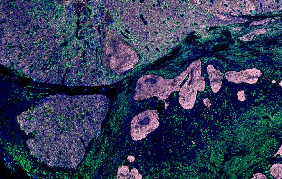
The new approach makes use of a cell or tissue’s own molecular makeup in order to better identify it. For example, the Michigan pill specifically targets breast cancer. So, in order to better identify issues, their pill focuses on “integrin.”
However, the entire body contains amounts of integrin — not just the breast. Despite this, Michigan researchers successfully located tumors as small as 2 cm in mice.
The Michigan pill works by using a target molecule that binds only with specific cell surface receptors. That means that the fluorophore will seek out cancer cells and “light them up”.
But since the pill is ingested, it needs to be absorbed into the bloodstream in order to work. The perks of injections are that you can pinpoint exactly where you want to identify potential malignant tissues and cells.
The molecular imaging approach works more effectively than injections, though.
It can provide more information about the type of tumor (benign, malignant, etc). In addition, tissue density does not affect this method nearly as much as other imaging techniques.
The pill still needs to be tested in humans, but its success could herald a new age in cancer screening, imaging, and diagnosis.


Comments (0)
Most Recent