
UC San Diego researchers have paved the way for alleviating over 15 different circulatory diseases. Using 3D printing, these nanoengineers successfully created a three-dimensional network of functional blood vessels with organic tissues.
One of the major obstacles to implanting organs produced by tissue engineering is replicating the functioning network of blood vessels that are needed to transport blood, nutrients, waste and other biological materials to and from the implanted tissue.
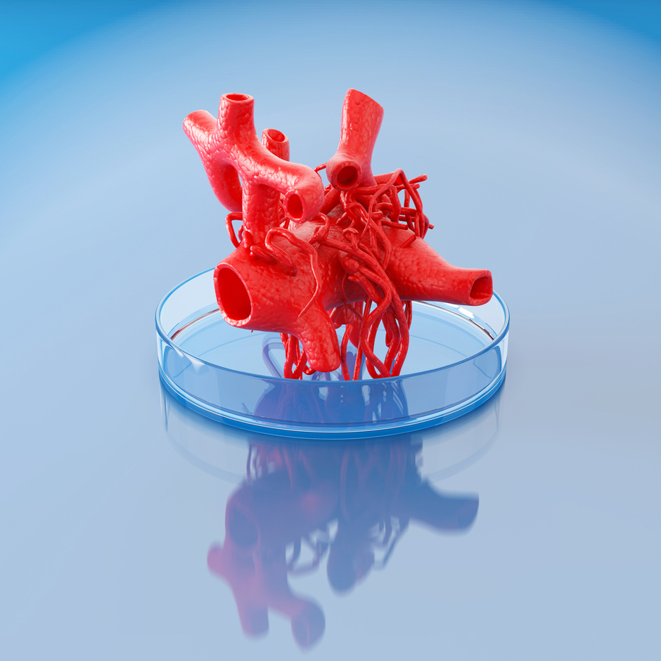
UC San Diego researchers successfully 3D printed organic vasculature.Click To TweetAlthough 3D printing techniques can now create dental or bone prostheses, for example, it’s not yet possible to print artificial organs due to the complexity of their micro-architecture. 3D printing living tissues has only worked in the smallest examples.
3D Printing Functional Vasculature
Led by Professor Shaochen Chen, a team of nanoengineers at the University of California San Diego 3D printed a network of biomimetic blood vessels. A paper on their work was published in the journal Biomaterials.
Lifelike and fully functional, these vessels can be safely implanted into the natural circulatory system via 3D printed organic tissues.
Professor Chen’s lab developed a bioprinting technology and used homemade 3D printers to produce intricate 3D microstructures that mimic natural blood vessels and can branch out into a network of smaller vessels.
Using 3D computer models, researchers can rapidly print one layer at a time, thereby creating a 3D solid polymeric scaffold, containing endothelial cells, the type of living cells that naturally form the inner lining of blood vessels.
This process uses affordable and biocompatible materials to print a detailed microstructure of vessels in high resolution, and in a matter of seconds, much faster than competing methods that take hours to print simple structures, usually a single blood vessel.
Chen’s Nanobiomaterials, Bioprinting, and Tissue Engineering Lab used the same technology to 3D print a liver tissue and microscopic “fish” which rummage about inside the tissue to spot and remove toxins.
UCSD researchers cultured some microtissues in vitro for one day before grafting them onto the skin wounds of mice. After examining the implants, they found that the microstructures had grown and merged with the host vasculature, circulating blood normally.
These blood vessels are not yet able to circulating nutrients and waste, but Chen and his team are working on a solution. They have already addressed one of the biggest challenges facing nano-bioengineering: vasculature, or the lack thereof. This achievement will lead to other advances in regenerative medicine.


Comments (0)
Most Recent