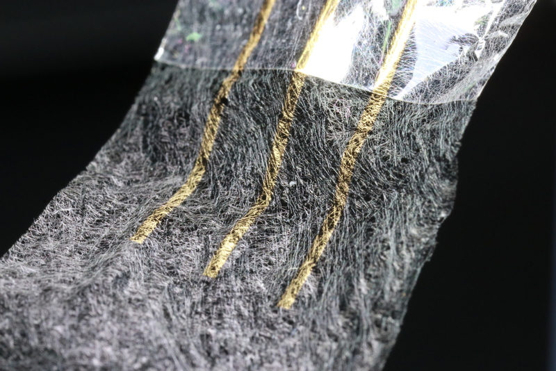
Japanese researchers have reportedly created a new heart sensor that can monitor cells with minimal disruption.
A team of researchers from the University of Tokyo and RIKEN in Japan created an electronic heart sensor that has minimal effect on the behavior of heart cells. During a demonstration, the team produced a working sample of heart cells attached to a soft nanomesh sensor, touching the tissues directly.
There is no doubt that the heart is one of the most significant life-sustaining organs in the human body. However, millions of people from around the world are suffering from cardiovascular ailments like arrhythmia, angina, and peripheral arterial disease. These common conditions make it impossible for many to live a normal life.
Now, researchers are conducting different experiments to find new and more effective treatments for these heart diseases. The new heart sensor created by Sunghoon Lee and his team could potentially be this sought-after treatment.
“When researchers study cardiomyocytes in action they culture them on hard petri dishes and attach rigid sensor probes. These impede the cells’ natural tendency to move as the sample beats, so observations do not reflect reality well,” Lee said.
“Our nanomesh sensor frees researchers to study cardiomyocytes and other cell cultures in a way more faithful to how they are in nature. The key is to use the sensor in conjunction with a flexible substrate, or base, for the cells to grow on.”
Read More: Google AI Predicts Heart Disease and Stroke From Retinal Images
The Nanomesh Heart Sensor
For their research, Lee and his team reportedly used the healthy culture of cardiomyocytes sourced from human stem cells. The team then placed this culture on top of a soft material known as fibrin gel. Then, the team placed the electronic heart sensor on top of the culture. They did so using a process involving adding and removing a liquid medium at a precise time.
“The polyurethane strands which underlie the entire mesh sensor are 10 times thinner than a human hair. It took a lot of practice and pushed my patience to its limit, but eventually I made some working prototypes,” Lee went on to say.

The sensors were created through electro-spinning which extrudes the ultrafine polyurethane strands into a flat sheet in a manner similar to how 3D printers work. The layer was then coated in a type of plastic called parylene to strengthen it. The team then removed the parylene on some areas of the sheet and replaced it with gold to make way for the sensor probes and communication wires.
The sensor has three probes which read the voltage present at three different places. These sensors allow researchers to see the propagation of signals that trigger the beating of the cells. This reading is highly significant in assessing the effects of drugs on the heart.
“Whether it’s for drug research, heart monitors, or to reduce animal testing, I can’t wait to see this device produced and used in the field. I still get a powerful feeling when I see the close-up images of those golden threads,” Lee was quoted as saying.


Comments (0)
Most Recent