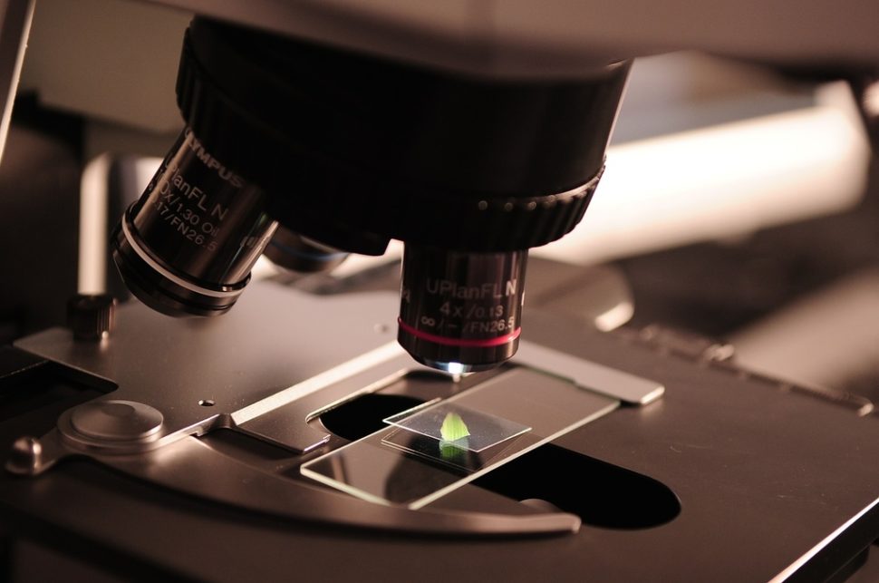
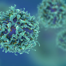
In another breakthrough in imaging technology, researchers were able to create a new hyperlens crystal so powerful it enables the viewing of ultra-small features on the surface of a living cell.
Just imagine using hyperlens to see virus-sized features on the surface of any living cell in its natural environment. Impressive, wouldn’t it be? While such imaging technology seems to exist only in science fiction films, a group of researchers was able to make it a reality.
In a paper published in the journal Nature Materials, researchers from Vanderbilt University in Tennessee reported how they constructed an instrument with such powerful capability. Apparently, a fundamental advance in the quality of a particular optical material made hyperlensing, or the process of creating lenses that can see objects way smaller than the wavelength of light, possible.
The said achievement in imaging technology could have a significant impact on biological and medical science and other research studies, especially those involved in the development of new antibiotics.
@VanderbiltU researchers developed a hyperlens to see ultra-small objects!Click To TweetDeveloping Hyperlens Crystal
The Vanderbilt researchers led by Professor Joshua Caldwell were able to create an optical lens powerful enough to see objects as small as 30 nanometers using the optical material hexagonal boron nitride (hBn), a natural crystal with hyperlensing properties.
For the record, the best previously recorded resolution using hBn was an object 36 times smaller than the infrared wavelength used. That’s about the size of the smallest bacteria known today.
Caldwell and his team made hBn crystal using isotopically purified boron which enhanced its potential imaging capability by about a factor of ten.
“We have demonstrated that the inherent efficiency limitations of hyperlenses can be overcome through isotopic engineering,” Alexander Giles, a research physicist at the U.S. Naval Research Laboratory and a member of the research team, said. “Controlling and manipulating light at nanoscale dimensions is notoriously difficult and inefficient. Our work provides a new path forward for the next generation of materials and devices.”
It should be noted that attempts to image living organisms using previous imaging devices with nanoscale resolution were unsuccessful because they require high-vacuum conditions or lethal sample preparation technique, an issue that the new hyperlens aims to address.
“Previously, I collaborated with another group to demonstrate that a flat slab of hBN could be used to image deeply sub-diffractional objects (~100 nm diameter) with long wavelengths of light (up to 13.2 um) using the hyperbolic phonon polariton modes within hBN,” Caldwell explained.
However, the professor said that it requires a scanning near-field optical microscope to read out the signal on the other side, which apparently was fine for proof of principle work only but not realistic for practical lenses. To make it work for practical lenses, he has to reduce the optical losses further.
“In this new paper, we were able to demonstrate that the losses in hBN could be reduced up to 14 fold, theoretically, with 3-4 fold reductions demonstrated experimentally by isotopic enrichment,” Caldwell went on to say.
“This allows us to support smaller wavelength modes at the same incident frequency of light, thus allowing higher spatial resolution imaging and the ability to propagate these smaller wavelength modes over longer distances in the hBN, which allows them to be collected on the other side.”
With the help of researchers from the University of California, San Diego, Kansas State University, Oak Ridge National Laboratory, and Columbia University, they were able to create a hyperlens crystal that could capture image features the size of a virus on the surface of a living organism in its natural environment.
Wondering how small that is?
To put it in perspective, an inch has 25 million nanometers, and a strand of human hair is about 80,000 to 100,000 nanometers in diameter. Our red blood cell is approximately 9,000 nanometers while viruses range from 20 to 400 nanometers only.
“Currently, we have been testing very small flakes of purified hBN,” said Caldwell. “We think that we will see even further improvements with larger crystals.”


Comments (0)
Least Recent