
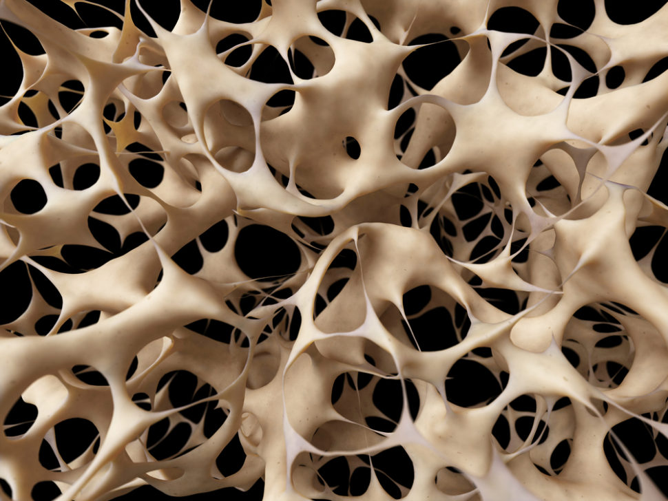
Researchers at Oregon Health & Science University have developed a material that can replicate human bone tissues and can be used to study bone diseases and injuries further.
The team noted how precise the “bone in a dish” replicated bone tissues, from its microscopic crystal structure to its biological activity.
“Essentially it is a miniaturized bone in a dish that we can produce in a matter of 72 hours or less,” Luiz Bertassoni, an assistant professor at the OHSU School of Dentistry, said.
“What is remarkable is that researchers in our field have become used to cultivating cells within a protein mixture to approximate how cells live in the body. But this is the first time anyone has been able to embed cells in minerals, which is what characterizes the bone tissue.”
The new material is highly significant in the study of bone functions, diseases, and bone regeneration. According to the researchers, it allows them to ask questions like “how bone cells attract different types of cancers; how cancer cells move into the bone; and how bone takes part in the regulation of marrow function.”
Studying Bone Diseases Through Synthetic Bone Tissues
The tissue-replicating material was created using a mix of human stem cells, and a solution filled with collagen, a protein found in the matrix of bone tissue. The mixture resulted in a gel embedded with stem cells.
Then, the researchers flooded the stem cell-filled gel with a mixture of dissolved calcium and phosphate, also known as the minerals of the bone. After that, Bertassoni and his team added the protein osteopontin that came from cow’s milk to prevent the minerals from forming crystals quickly.
The final mixture was diffused through a network of channels, whose width is about the size of a DNA strand, in the spongey collagen. Then, the dissolved minerals precipitated into organized layers of crystals.
“We can reproduce the architecture of bone down to a nanometer scale,” Bertassoni added. “Our model goes through the same biophysical process of formation that bone does.”
In the said environment, the bone cells osteoblast and osteocytes were developed without any other molecules, acting like they’re in an actual bone matrix.
Within a few days, the living cells squeezed the narrow protrusions through the spaces in their mineralized environment, allowing them to communicate and connect with adjacent cells.
The team tested their lab-made material on laboratory mice, implanting it beneath the rodents’ skin. The lab-grown blood vessels had connected to the vasculature of the rodents’ bodies after a few days.
When the mice were injected with prostate cancer cells, the researchers observed that tumor growth had increased three times more in the mice with mineralized bones than those with natural bones.
Bertassoni and his team are now working on a version of the material that contains marrow cells within the surroundings of artificial bone for studying the beginning and development of blood cancers.


Comments (0)
Most Recent