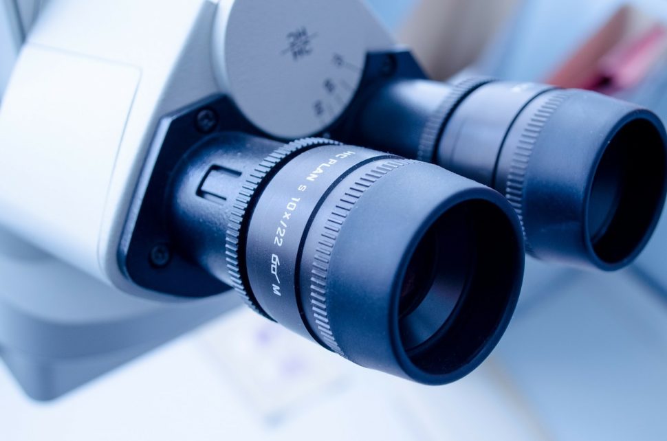
Scientists have invented the DNA microscopy to provide a completely new image of cells.
In the past, we depended on light, x-ray, and electron to give us a glimpse of the tissue and cell. But the technology got a bit more advanced.
Now we have a clear view of happenings in the thread-like fibers in the brain, including how embryos develop into rudimentary organs in the mouse.
But, as advanced as microscopy has gotten, one major flaw still exists; our inability to see cell activities at the genomic level. That’s about to change.
Biophysicist, Joshua Weinstein, and his colleagues have invented a new type of imaging that provides genomic level images. And they’re calling it DNA microscopy.
That’s because the new microscope uses DNA bar codes – rather than light or any other optics- to find the exact position of molecules within a sample.
With this new imaging technique, scientists can create an accurate picture of cells and collect extensive genomic information at the same time. In other words, it provides a layer of biology that has remained unexplored until now.
Speaking on the new invention, one of the Weinstein’s colleagues and Howard Hughes Medical Institute (HHMI) investigator, Aviv Regev said:
“It’s an entirely new category of microscopy. Not only is the technique new, but it’s a way of doing things that we haven’t ever considered doing before.”
So, how does the technique work?
How DNA Microscopy Works
To capture an image of cell activities at the genomic level, first, the scientist fixed lab-grown cells into position in a reaction chamber.
Next, they added an assortment of DNA bar codes to stick with the RNA molecules. That way, each of them would end up with a unique tag.
Using a type of chemical reaction, the scientists created more copies of these tagged molecules. This, in turn, caused the molecules to expand out from their original location.
According to Weinstein, it’s like a radio tower broadcasting its signal outwards.
At some point during the process, the tagged molecules collided with each other and linked up into a pair. While closer molecules collided to generate more DNA pairs, those further apart produced fewer pairs.
In a 30-hour process, the team spelled out the letters of every molecule in the sample using a DNA-sequencing machine. A previously created algorithm then decoded the data and converted it into images.
Weinstein noted:
“You’re basically able to reconstruct exactly what you see under a light microscope.”
According to the researcher, both methods are complementary.
Light microscopy provides a better image of the molecules when they’re sparsed within a sample. DNA microcopy, On the other hand, excels when the particles are tightly-packed – even piled on top of each other.
Weinstein believes that this new form of microscopy could one day let scientists deliver faster immunotherapy treatment. That means, cancer patients would have a better fighting chance.
“The method could potentially identify the immune cells best suited to target a particular cancer cell,” he says.


Comments (0)
Most Recent